How fearful events are represented in the hippocampus, the brain’s memory center, is still elusive. In a new paper published in Nature Communications, Albert Barth and Marta Jelitai from Viktor Varga’s lab at the HUN-REN Institute of Experimental Medicine (HUN-REN IEM) described aversive stimulus-triggered single-neuron and population responses, as well as alterations of the spatial code in the dorsal hippocampal CA1 region.
As demonstrated by the Nobel Prize-winning discovery of John O’Keefe and Jonathan Dostrovsky, about 30-50% of hippocampal principal neurons, also known as pyramidal cells, exhibit position-dependent activity (these neurons termed place cells). Therefore, Barth and Jelitai had to separate spatially modulated activity from the response to the aversive stimulus by varying the location of the air puff, the fearful stimulus, while monitoring the activity of dozens or even more than a hundred dorsal hippocampal neurons.
In case of some neurons, the air puff-evoked dramatic increase of activity was obvious even by looking at the raw recordings: a near silent neuron started firing up to 50 action potentials per second as the intense airflow reached the mouse’s face. When the location of the air puff was changed, the neuron “followed” the stimulus and stopped firing at the original location. Thus, these neurons were not sensitive to the animals’ whereabouts but were rather tuned to the aversive event. In contrast, those principal neurons that preferred to fire at the stimulus location before the aversive event were suppressed whereas place field of other place cells shifted to other locations.
Most neurons work together for representing the outside world or the inner state of the animal by a population code composed of their coordinated activity. Thus, Barth, Jelitai and their colleagues had set out for deciphering the population code of fearful stimuli and managed to identify neural patterns tuned for aversive events. Strikingly, they could detect these patterns even before the animal had encountered the air puff for the first time. This latter finding suggests that certain hippocampal neurons are sensitive to the co-occurrence of stimuli signaling imminent dangers, such as intense sound and light of an explosion.
One consequence of population coding is the possibility of estimating the animal’s position based on the coordinated activity of simultaneously registered place cells. The authors asked how this place code had been affected by the aversive event. This analysis has brought the most unexpected discovery: the stimulation distorted the position code, but not randomly. Instead of representing the current location where the animal was exposed to the air puff, the activity of co-registered neurons pointed to the location of the reward, as if the mouse had started thinking about a recent pleasant experience at the time of the adverse event.
These discoveries raise multiple questions, the answers to which may pave the way for clinically relevant innovations. Would it be possible to selectively manipulate these aversive event-coding neurons? What would happen if their activation was prevented? Could the formation of negative memories, or even the onset of traumatic event-triggered pathological mental states, be prevented or treated?
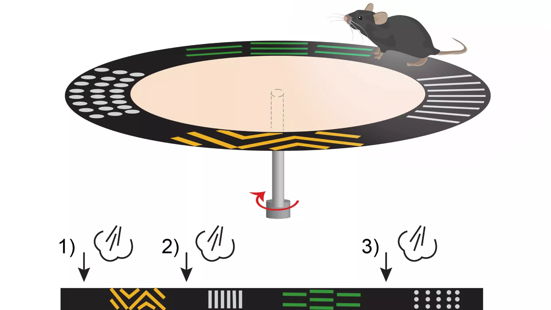
Schematic diagram of the experiment. The pattern on the plate helps to identify the activity of the different site cells. The location of the air puffs is indicated by the arrows in the lower figure.