Using state-of-the-art neuroanatomical, physiological and computational cell modelling techniques, a research team led by Gábor Nyiri at the HUN-REN Institute of Experimental Medicine (HUN-REN IEM) has created the most detailed models to date of two important cortical inhibitory neurons. The models can be built on using further experimental data and are therefore excellent for interpreting the results of physiological experiments, for studying the role of the hippocampus in learning and memory processes, and for creating neural network models that simulate its function. The paper was published in the prestigious journal PLoS Biology with Virág Takács, Zsuzsanna Bardóczi, Áron Orosz, Ábel Major, and Luca Tar as joint first authors.
Nerve cells are basically made up of three different parts: the cell body, the dendrite and the axon. The dendritic tree, which branches like a tree branch, receives information (inputs) from other nerve cells. The axonal tree, made up of long branches, transmits the nerve cell's impulses (output), most often to the dendrites or cell bodies of other nerve cells. Neural transmission goes through special junctions between two cells, called synapses.
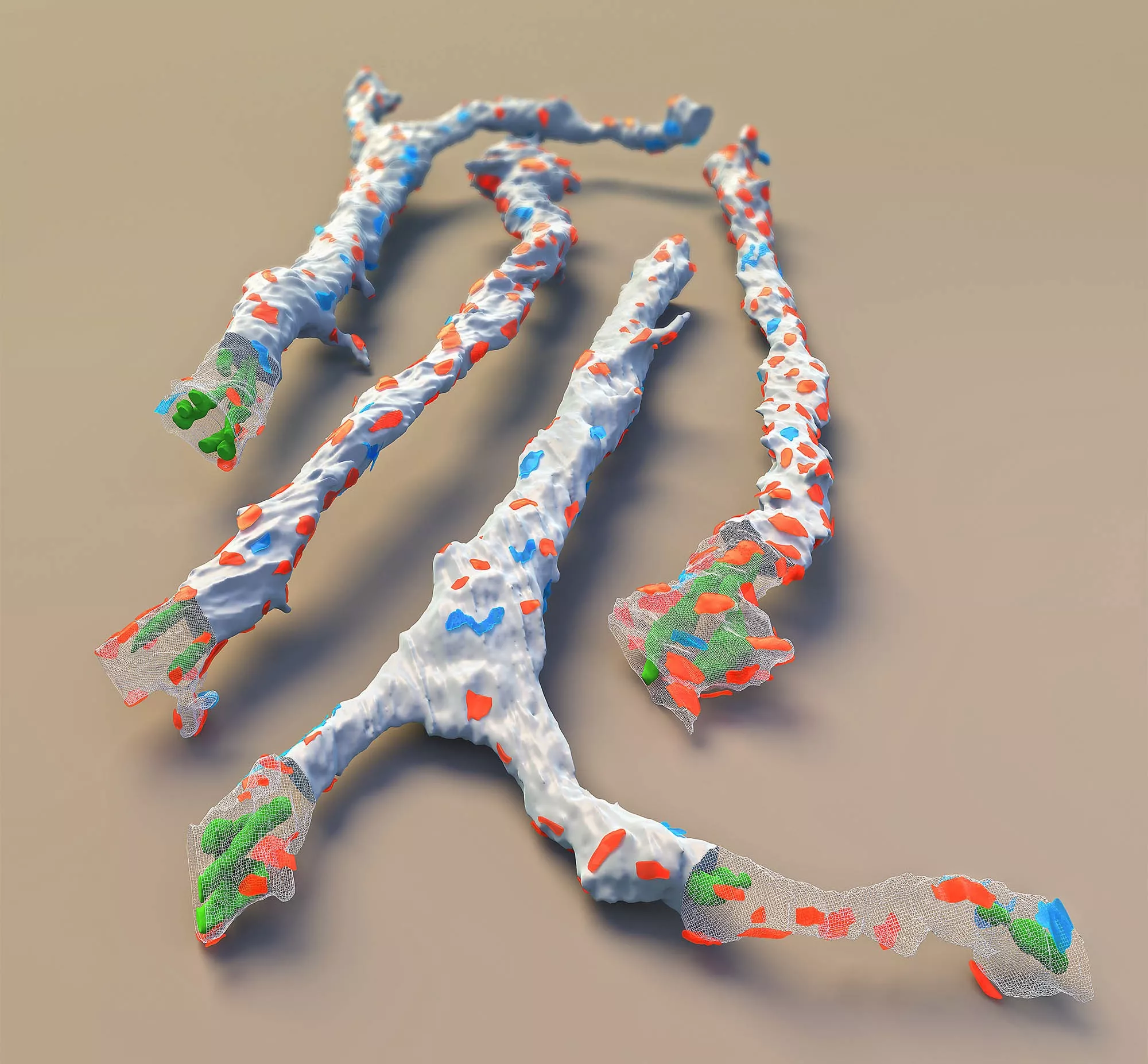
Inhibitory neuron dendrite segments (four examples) with mitochondria (green) inside and inhibitory (blue) and excitatory (red) synapses located on their surface.
The hippocampus is a cortical area of fundamental importance for the formation of memory traces, where a variety of morphologically distinct inhibitory interneurons, responsible for different tasks, regulate the function of the excitatory pyramidal neurons. Gábor Nyiri's group investigated two important representatives of somatostatin-containing inhibitory interneurons that have different network functions. The Oriens-Lacunosum Moleculare (OLM) cells innervate the dendritic tree of the pyramidal cells furthest from the cell body, while Hippocampo-Septalis (HS) cells have, in addition to their local axon branches, fibres leaving the hippocampus and reaching several other distant brain areas (mainly the medial septum).
The spatial dimensions of different cellular components, such as the diameters of different dendrites, the volume of cytoplasm inside them, the surface area of membranes, and the number, ratio and size of excitatory and inhibitory synapses on dendrites are very important parameters for the propagation of excitation within the cell. These can be used to create computational models simulating the functioning of individual cells and neural networks. However, due to the lack of appropriate testing methods and the labour-intensive nature of measurements, researchers have not yet had precise data on the morphological characteristics of interneurons in the hippocampus.
Gábor Nyiri's group described the morphological parameters of OLM and HS cells with high accuracy using a state-of-the-art scanning electron microscope at HUN-REN KOKI. The first essential step of the work was to set up the new instrument and the appropriate sample preparation techniques. The two cell types and their different types of synapses were specifically labelled, and their cell bodies, different sections of dendrites and axons were reconstructed in three dimensions by taking serial electron microscopy sections using several software tools.
The team's results show that although the dendrites of the two cell types are similar in their location and light microscopic appearance, their electron microscopic anatomy ‒ and thus their function ‒ differ significantly. It has been shown that the dendrites and cell bodies of HS cells are much more densely covered by synapses, so that the number of inputs per the total cell surface is almost twice that of OLM cells (15,600 vs. 8,400, respectively). They also measured the basic physiological properties of the two cell types and combined these with anatomical properties and literature data to produce the most detailed and anatomically accurate 'passive and active' interneuron models to date. These can be used to incorporate further experimental data in the future and are therefore excellent tools for interpreting the results of physiological experiments, studying the role of the hippocampus in learning and memory processes, and creating neural network models simulating its functioning.