Researchers from the ELKH Institute of Experimental Medicine (IEM) and the ELKH Research Centre for Natural Sciences (TTK), also affiliated with the University of Veterinary Medicine have uncovered new genetic features of the brain's reward system. The research, carried out by Ferenc Mátyás, head of the Neuronal Networks and Behaviour Research Group, and his PhD student Ákos Babiczky, focused on the anatomical connections between the so-called medial prefrontal cortex (mPFC) and two ancient regions of the central nervous system, the striatum and the tegmentum, in mice. The latter two form the pathway where dopamine – the 'reward molecule' – orchestrates learning and other reward-related processes. A study presenting the results of the research has been published in the prestigious international journal eLife.
Although this system has been intensively investigated in the previous decades, certain molecular features were not completely elucidated. Unfortunately, without a precise anatomical framework, functional investigations cannot be carried out reliably. For example, it was not clear whether a single neuron of the mPFC could simultaneously innervate the striatum and the tegmentum.
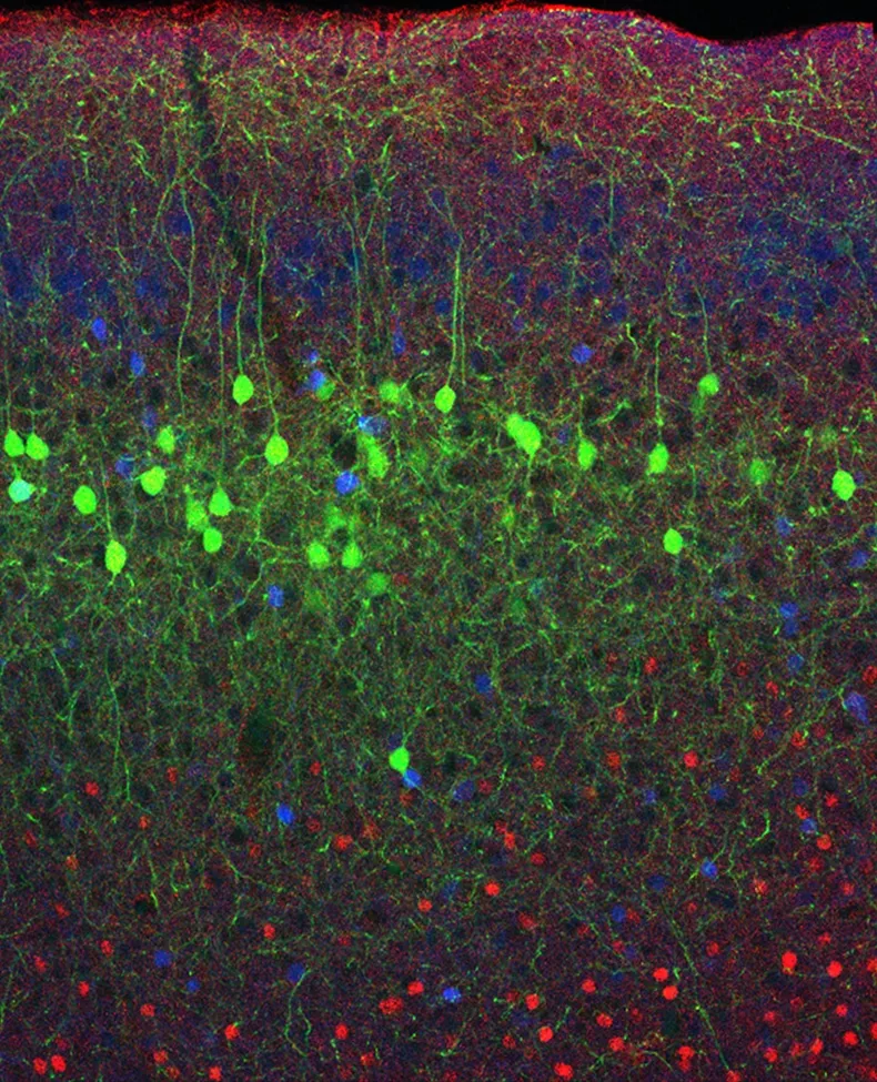
different populations labelled with red and blue. The method is reliably applicable to differentiate distinct neuron populations.
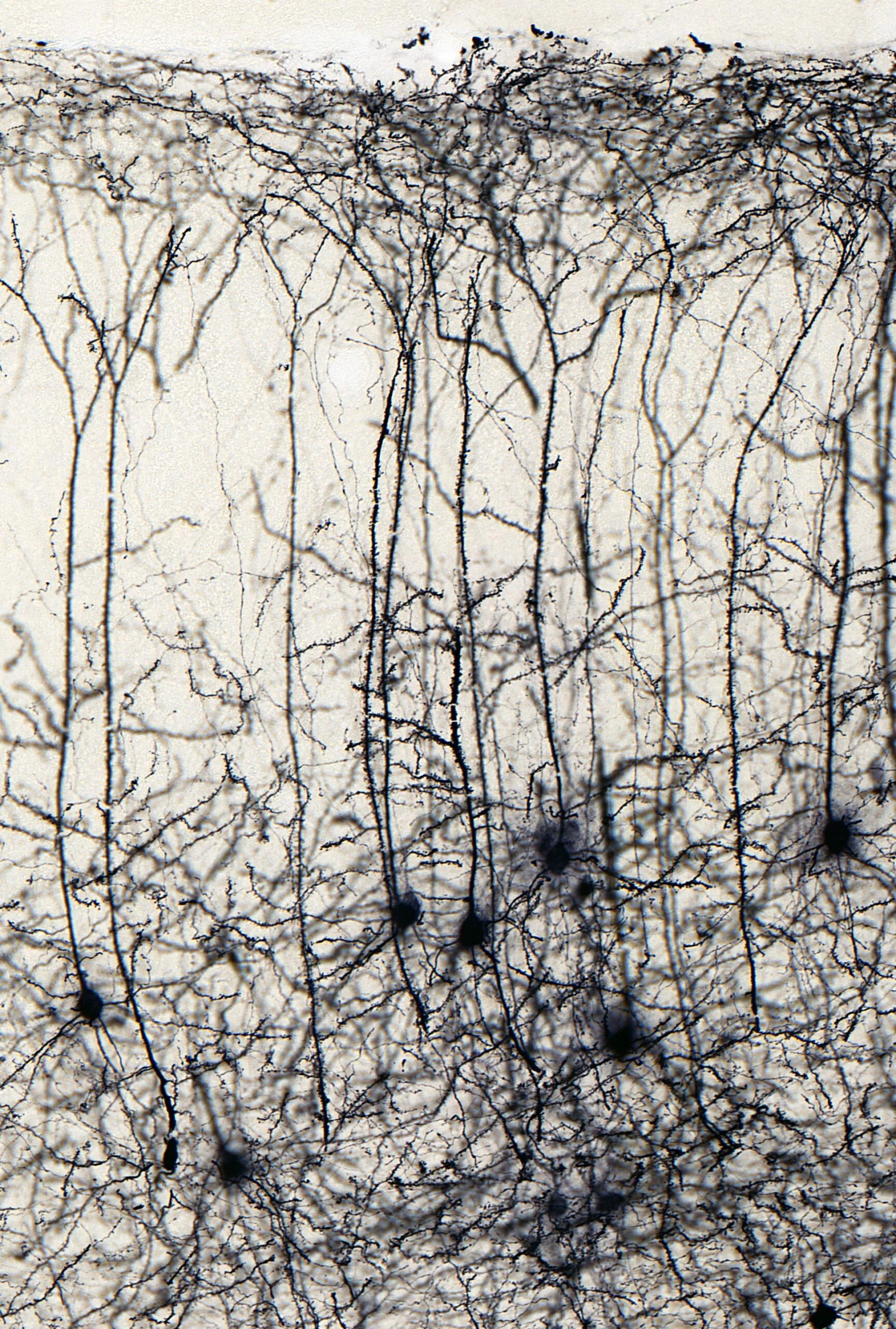
In order to clarify this, first, the researchers described where striatum- and tegmentum-innervating neurons are localized in the mPFC, using fluorescent microscopy, several genetically modified mouse strains and special viral constructs. The results clearly showed that the two populations occupy different layers and subregions in the mPFC and they also revealed that these neurons have different genetic identities. Next, they directly compared them and found that there is minimal overlap between the two populations indeed. Furthermore, the researchers also demonstrated that these two populations have different connections with other brain regions as well.
throughout the whole brain.
These new results implicate that the mPFC – widely known as one of the most important higher order regulators of behavior and personality – can modulate the activity of the striatum and the tegmentum independently. This can be directly tested in the future, because the experimental approach described by the authors of this study published in the journal eLife can easily be applied to physiological and behavioral experiments. Untangling these connections at the anatomical and functional level can help us understand the healthy operation of the reward system as well as its malfunctions, like addiction.
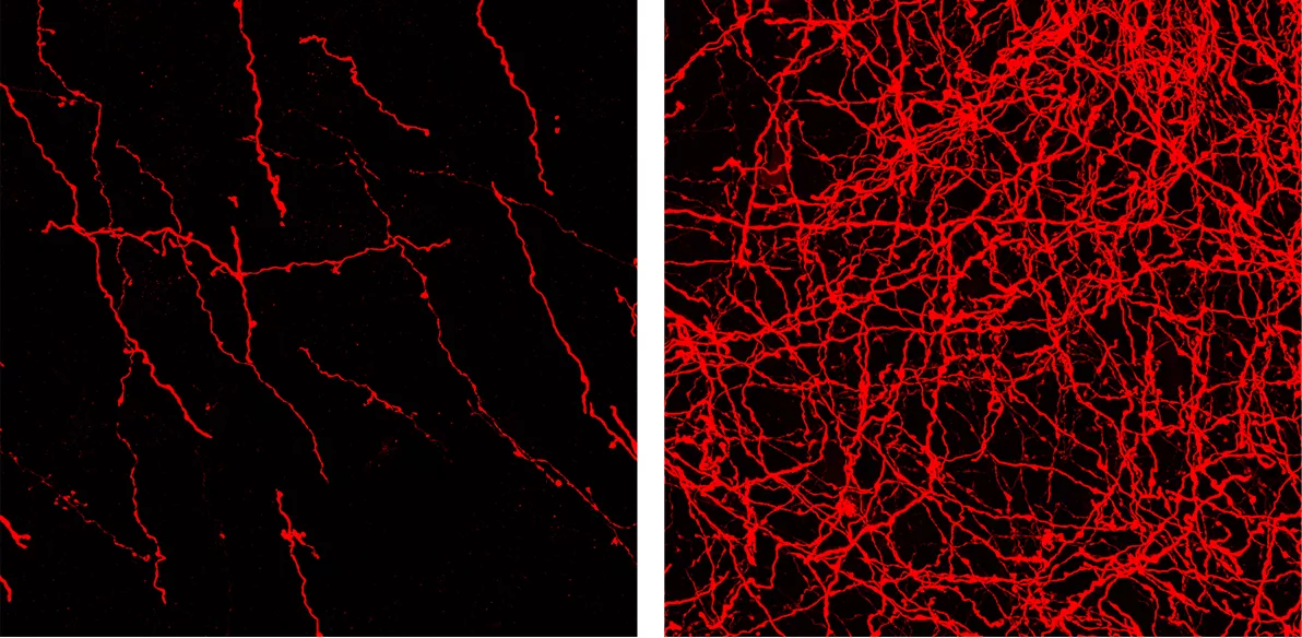
axons are visibly different in the two regions.
Publication:
Ákos Babiczky, Ferenc Mátyás (2022). Molecular characteristics and laminar distribution of prefrontal neurons projecting to the mesolimbic system. eLife. doi: 10.7554/eLife.78813