In their new seminal study, researchers of the ELKH Institute of Experimental Medicine (IEM), the Pázmány Péter Catholic University (PPCU) and the BrainVisionCenter (BVC, founded in 2021 by Botond Roska, Balázs Rózsa and ITM), in collaboration with their colleagues at Washington University in St. Louis identified and described a new functional principle that is important in the early phase of learning and that is present uniformly in the whole cerebral cortex. The paper was published in the prestigious international journal eLife.
The cortex represents the outside world in the form of internal models that help predict important external events and direct behavior. Therefore, these representations have to contain information about the value of these events, meaning if they are beneficial or potentially dangerous. It is still unknown how this information integrates into the relevant representations. The present study aims to show such a mechanism on the local cortical circuit’s level. Using a previously unavailable tool and method the researchers recorded the activity of an important but scarcely present interneuron, the VIP-type (vasoactive intestinal polypeptide-expressing) interneuron. This inhibitory interneuron type represents barely 1% of the cortical cell population, yet it is very important in modulating the activity of the local cortical circuits through a powerful mechanism called disinhibition. VIP neurons do inhibit other inhibitory interneurons that suppress the activity of the pyramidal cells, thereby modulating the input gain on these neurons, that may provide a powerful tool in the learning process.
Due to the limitation of the methods available up to date (electrophysiology and imaging) our knowledge of this very important cell type is limited too. The reach of these methods misses the cell types that are rare in the cortex. The new tool, developed in the laboratory of Balázs Rózsa overcomes this limitation: the 3D acousto-optical scanning 2p microscope is able to record the chosen cells within a large cortical volume, even if they are sparse and have a small cell body. Recording speed, spatial resolution and signal-to-noise ratio of the special microscope far outperforms the imaging tools used so far. Additionally, population activity of the sparse neurons even in deeper-laying regions like the medial prefrontal and the auditory cortices were recorded in the laboratory of Adam Kepecs by fiber photometry.
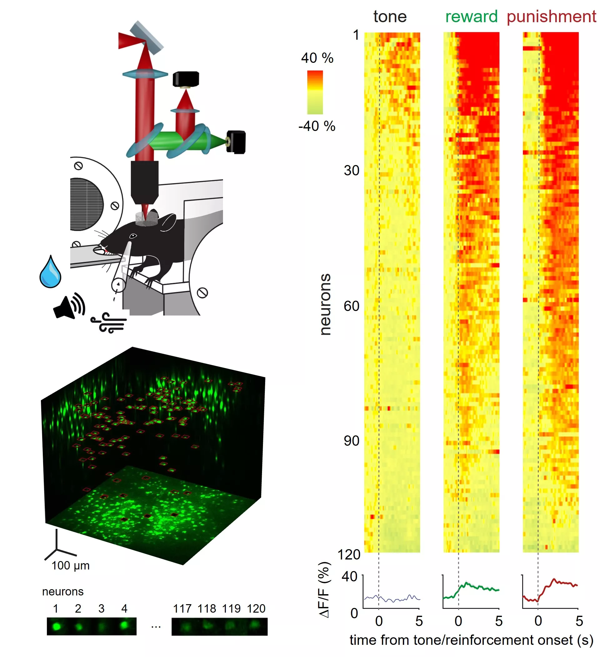
Figure 1: Experimental setup and functioning of VIP interneurons. Mice had to discriminate two different tones and got rewarded or “punished” according to performance, whilst the activity of the VIP interneurons were recorded by a 3D laser scanning microscope in a cortical volume of half a cubic millimeter. On the right panel: the activity of 120 VIP neurons, a neuron per row, time on the x axis. Red colour means activity, dashed lines tone and outcome onset. To the tone alone, only a subset of the cells is responsive, at the same time to either the reward or punishment most of the cells respond.
The protocol of the experiments that was conducted by Zoltán Szadai, Hyun-Jae Pi and Quentin Chevy with the technical assistance of Lidia Popara consisted of controlled-water-access mice discriminating two tones. Successfully performing mice were rewarded with drops of water, whilst an error led to an aversive air puff. To some surprise both the reward and the “punishment” led to a strong, phasic activation of the VIP cells, and all over the cortex. This outcome shows that VIP interneurons actually relay information locally about an organism-level information on reinforcers, helping regulate local processing and leading to plasticity.
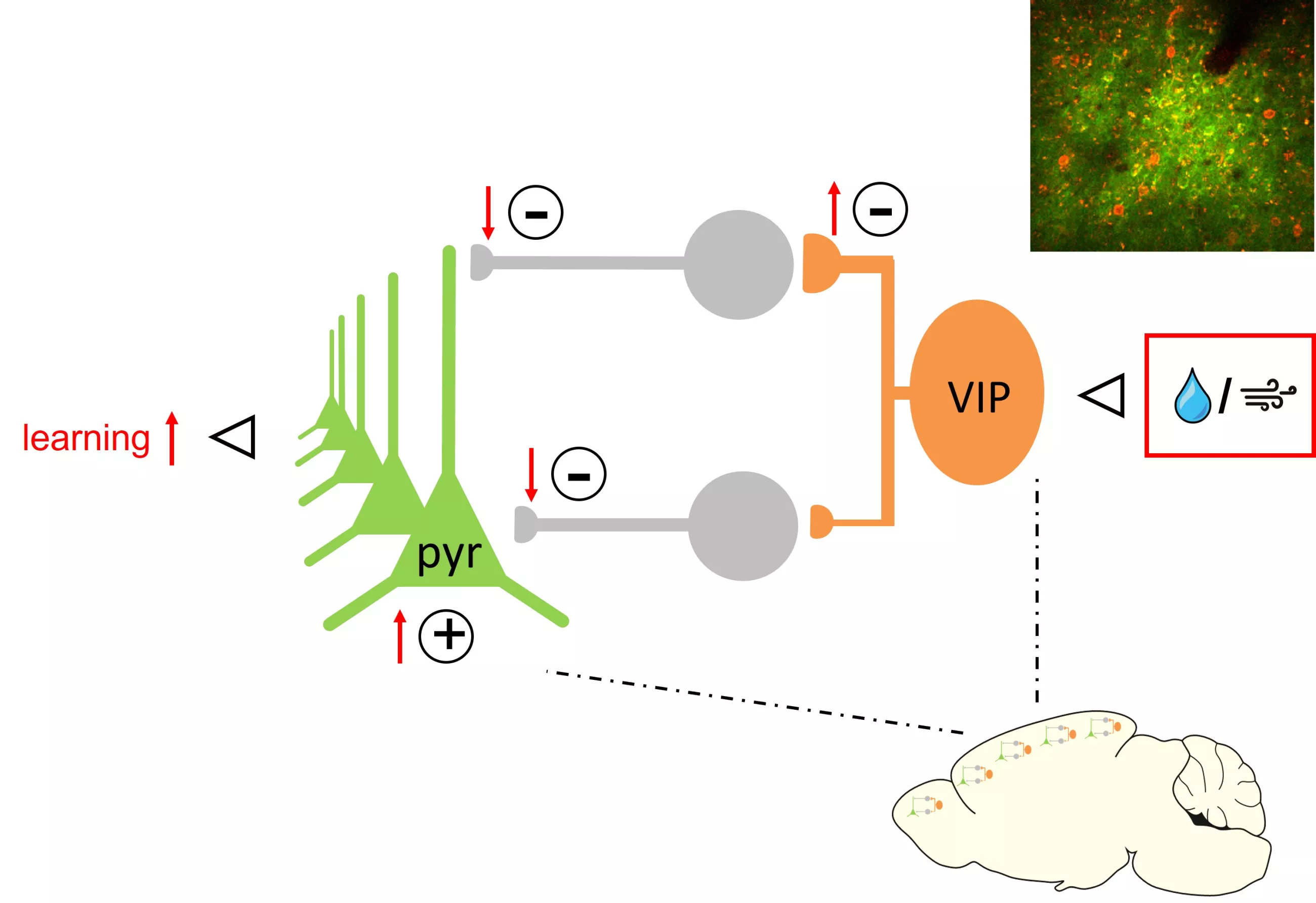
Figure 2: Functioning of cortical microcircuits: in response to either reward or punishment, VIP cells increase their activity therefore inhibition of the inhibitory cells. In return the activity of the pyramidal cells increases. Plus and minus signs designate activating or inhibitory effect on the postsynaptic neuron. Red shows change in response to reward/punishment. Top right panel shows red-labelled VIP cells being scattered in the green cloud of pyramidal cells.
The study thus represents a step closer to understanding a cortex-wide mechanism of perception and learning, therefore provides basis for further investigations of healthy and pathological changes. The study also provides evidence for a similarity of reinforcement learning in organic brains and artificial intelligence models.
Publication:
Zoltán Szadai, Hyun-Jae Pi, Quentin Chevy, Katalin Ócsai, Florin D. Albeanu, Balázs Chiovini, Gergely Szalay, Adam Kepecs, Balázs Rózsa (2022). Cortex-wide response mode of VIP-expressing inhibitory neurons by reward and punishment. eLife, 11. Doi: 10.7554/eLife.78815.