In their new seminal study, researchers of the Thalamus Research Group led by Dr. László Acsády of the ELKH Institute of Experimental Medicine (IEM) in collaboration with Lausanne University describe a new top-down communication channel between the cortex and its most important subcortical partner in information processing called thalamus. This region-specific new neuronal pathway is present in frontal cortical regions which are involved in higher order cognitive functions, but it is absent in parietal regions which deals with sensory information. The data demonstrate that the principles of information processing display regional heterogeneity in distinct cortical loops. The paper was published in the most prestigious neuroscience journal Nature Neuroscience.
The cortex is at the apex of information processing in the mammalian brain. Interestingly, however, beside olfactory inputs, no fast, precise information reaches the cortex without a thalamic transfer. Indeed, without exception all cortical regions receive thalamic inputs and none of them (olfactory cortex included) is functional without intact thalamic inputs. The interaction between the thalamus and cortex is not unidirectional. Every cortical region not only receives but also sends nerve fibers to the thalamus. This top-down information channel is called corticothalamic pathway. Thus, information between thalamus and cortex does not simply travel one way, rather it is processed in complex perpetually interacting cortico-thalamo-cortical loops. The new study by Nóra Hadinger et al. describes a novel corticothalamic connection.
Connectivity between cortex and thalamus has been intensively studied in the past 100 years and has been regarded canonical. This means that it was thought that there were no qualitative regional differences in the organization of the corticothalamic pathways despite the fact that there are obvious differences in the precise nature of information processed in various cortical areas (e.g. sensory, motor, emotional, etc.). In their new study Hádinger et al. show that the frontal cortical regions display a specific corticothalamic innervation pattern which is absent in other cortical areas demonstrating that the corticothalamic pathway is non-canonical. The new region selective corticothalamic pathway targeted the thalamic reticular nucleus (TRN) which contains only inhibitory neurons.
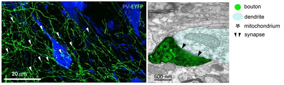
Figure 1: L5 collaterals originating from the frontal cortex tufted with synaptic terminals in the TRN. Left: Confocal image of L5 collaterals bearing synaptic terminals (EYFP+, green) in the TRN (TRN cells are labelled by parvalbumin ‒ PV, blue immunostaining). Arrows point to synaptic terminals. Right: Electron microscopy image of a L5 synaptic terminal (labelled by black precipitate and shaded in green) in the TRN (the postsynaptic TRN dendrite is shaded in blue).
There is no proper neuronal activity in the brain without precisely organized inhibitory activity. In the vast majority of diseases affecting the brain the balance and timing of excitation and inhibition is perturbed. Thalamus is not an exception. The main controller of thalamic inhibition is TRN. Inhibitory axon terminals of TRN densely innervate all thalamic nuclei. What controls TRN, controls thalamus as well. TRN forms a thin shell around the thalamus and its distinct sectors project to different thalamic regions in a nice topographic order. Since specific thalamic nuclei project to specific cortical regions,
activation of a specific TRN sector will result in governing the activity in a well-defined cortico-thalamo-cortical loop. And this is exactly what Hádinger et al. found. TRN cells targeted by frontal cortical inputs innervated those parts of the thalamus which projected back to frontal cortex closing the cortico-thalamo-cortical loop.
Inputs may be weak or strong, few or numerous. According to Hádinger et al. TRN receives many corticothalamic inputs, sometimes even from different cortical regions. The data also shows that TRN is especially sensitive to the synchronicity of its cortical inputs. Increasing cortical synchrony will gradually alter the timing and amount of TRN activity. This will be pivotal to control the response of thalamic cells to synchronous or hypersynchronous cortical activity, like epileptic seizures.
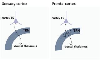
Figure 2: Schematic figure showing cortical L5-thalamus connection in the sensory and frontal cortices
In summary, the data suggest that the properties of information processing in cortico-thalamo-cortical loops involved in higher order cognitive functions are qualitatively different from those of other (e.g. sensory) loops. The new corticothalamic pathway targeting TRN will help to understand the neuronal basis of normal cognition and chronic neurological and neuropsychiatric diseases linked to frontal cortex including Parkinson’s disease, schizophrenia, chronic pain, and epilepsy, and it opens up novel avenues to study corticothalamic interactions.
The research was supported by ELKH and by the European Research Council within the framework of the Horizon program.
Publication:
Nóra Hádinger, Emília Bősz, Boglárka Tóth, Gil Vantomme, Anita Lüthi & László Acsády (2022). Region-selective control of the thalamic reticular nucleus via cortical layer 5 pyramidal cells. Nature Neuroscience. Doi: 10.1038/s41593-022-01217-z